Blender – facilitation the segmentation of coronary vessels
Challenge: Coronary vessel segmentation
Coronary vessel segmentation is a difficult process. Firstly, the resolution of a CT image is low when compared to the vessel diameter. For the tips, we have as many as several points per diameter. Secondly, the acquisition of the beating heart requires a complicated gating procedure which may result in artifacts in certain cases. Thirdly, in many cases there is relatively high noise in the image. Fourthly, the calcified sites have a similar radiographic density expressed in Hounsfield units and the differentiation between the plaque and the vessel lumen can be made solely based on the object shape.
Consequently, the manual segmentation process must comprise partial geometry reconstruction based both on the image and on the knowledge of anatomy of the segmenting person. The standard segmentation software usually offers limited 3D modelling methods, while the so-called 3D modellers do not enable to inspect a CT image during mesh modification.
What we did
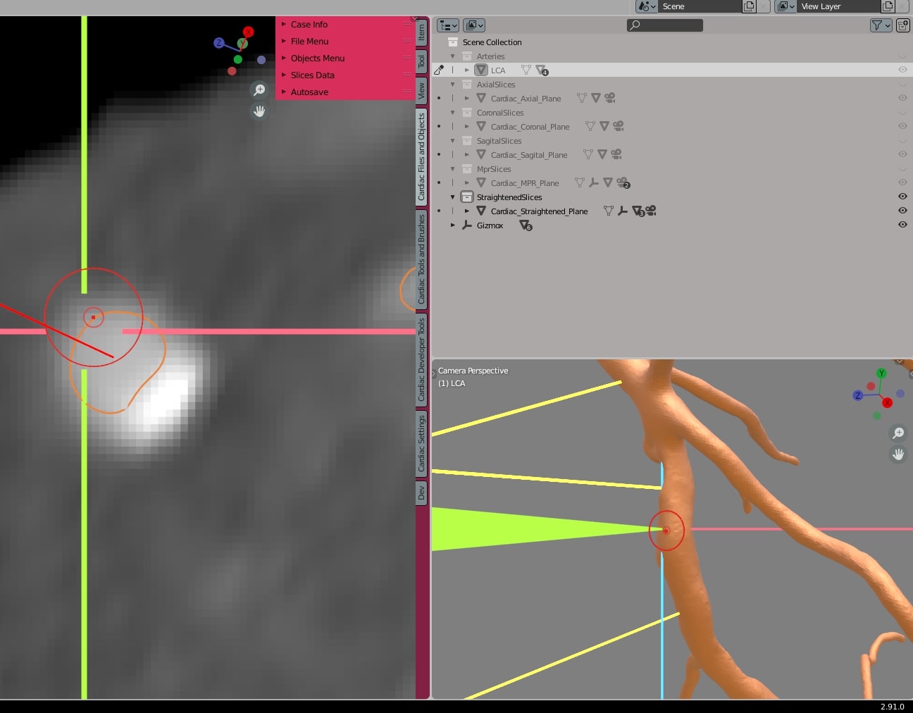
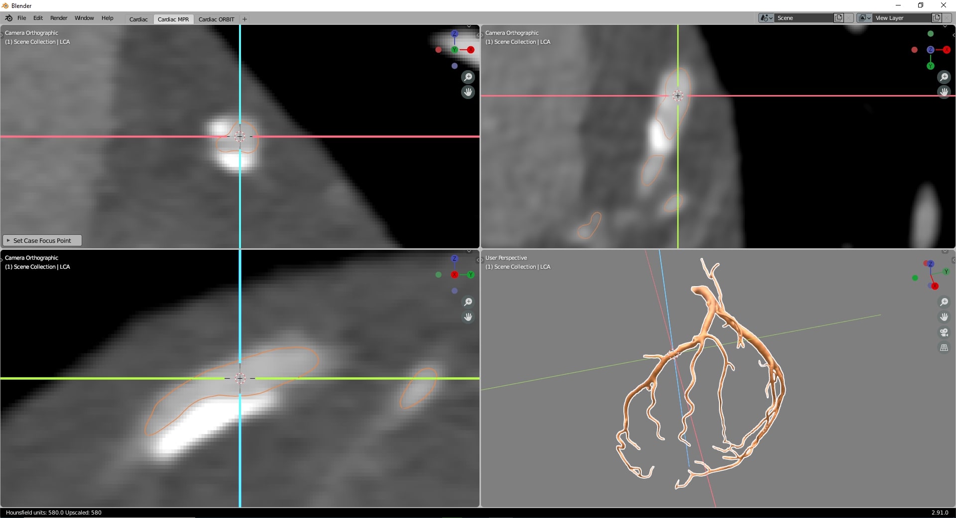
Having carried out the analysis, we decided to use Blender and provide this software with the required plug-ins for inspecting coronary vessels in MPR and CPR views. What is more, such an approach allowed us to implement the connection to the image storage and annotation system which highly reduces possible errors when handling hundreds of exams.
Results of our work
Thanks to the approach adopted by us, we obtained a tool which facilitates the segmentation of coronary vessels from CT significantly. It enables to inspect different DICOM views precisely and also, based e.g. on the generated coronary vessel cross-sections, allows to match the diameters to the contrast-filled artery areas fast. Our Blender plugin helps to update the outlines on an ongoing basis which provides the possibility to verify the introduced changes fast.

References:
