Cardiovascular image analysis: AI and more
At Graylight Imaging, cardiovascular image analysis lies at the very center of our interest. As you may remember, a few weeks ago, we announced our participation in the European Congress of Cardiology. This major event attracted over 31,800 attendees, including 5,400 faculty and presenters, and representatives from various cardiac societies and industries. We were excited to showcase two of our innovative works at this prestigious gathering. While we’ve briefly touched upon these cardiac image processing projects before, we’d like to now provide a more in-depth look at each.

Automating cardiac image analysis
Cardiovascular image analysis has seen significant advancements in recent years, enabling automated quantification of various cardiac parameters. One such application lies in determining the coronary artery calcification (CAC) score, a crucial metric for assessing future cardiac events. But not only.
The CAC score influences the treatment and the decision between conservative medical therapy, percutaneous coronary intervention (PCI), or coronary artery bypass grafting (CABG). What’s more, it can provide valuable insights into long-term outcomes after PCI, aiding in patient counseling and follow-up care planning. Finally, a higher CAC score can predict a more challenging PCI procedure. This knowledge enables interventional cardiologists to anticipate potential difficulties and select appropriate techniques and devices.
Coronary artery calcification quantification using cardiac computed tomography (CT) is a routine clinical practice. However, the current standard of care involves acquiring both contrast-enhanced and non-contrast CT scans, leading to increased radiation exposure for patients. We decided to address this limitation.
Advanced techniques for cardiovascular image analysis
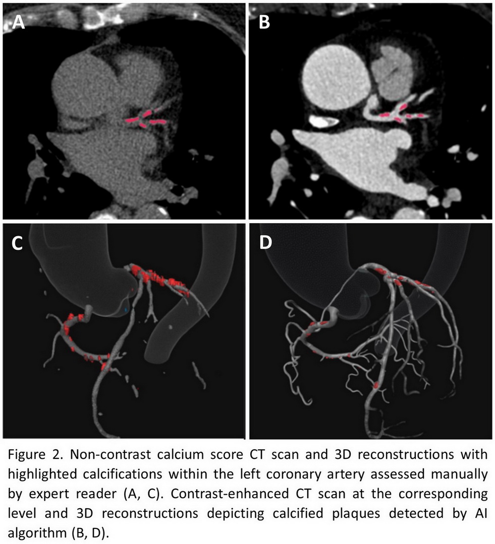
W developed a novel artificial intelligence (AI)-based algorithm capable of accurately determining CAC score solely from contrast-enhanced CT scans. Our approach leverages advanced deep learning techniques to extract meaningful features from the images, enabling reliable segmentation and quantification of coronary calcifications.
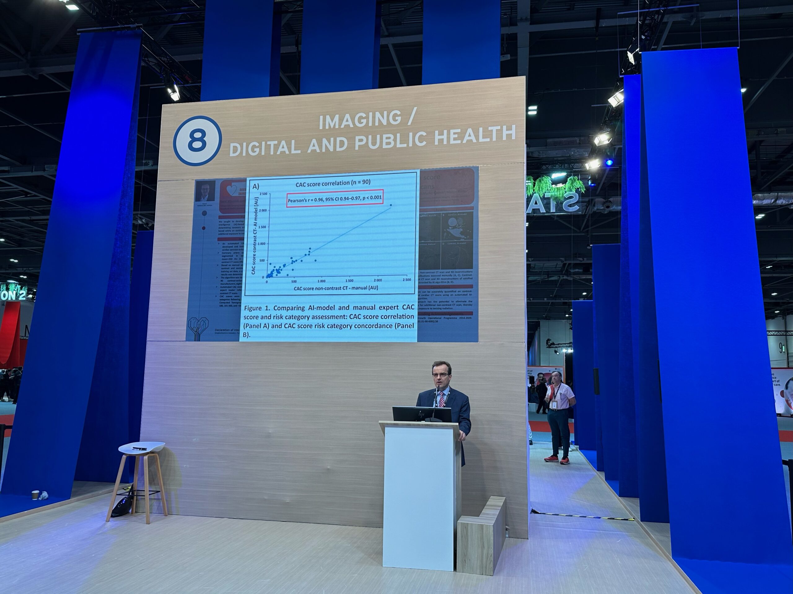
The algorithm was trained on 297 cardiac CT studies, encompassing a diverse range of patient demographics and clinical presentations. Additionally, we incorporated data augmentation techniques to artificially expand the training set and mitigate overfitting. In our evaluation, the AI-based algorithm demonstrated exceptional performance, achieving a high correlation with manual reference CAC scores. Furthermore, the model consistently classified patients into the correct CAC risk categories, indicating its clinical utility for risk stratification.

Reducing the patient’s exposure to ionizing radiation
By eliminating the need for non-contrast CT scans, our AI-based approach offers a significant advantage in terms of radiation dose reduction. This is particularly important for patients undergoing multiple cardiac CT examinations, such as those with ongoing cardiovascular disease or those at high risk for future events.
In conclusion, we hope that our study presents a promising AI-based solution for CAC score quantification in contrast-enhanced cardiac CT. By reducing radiation exposure without compromising accuracy, this advancement in cardiovascular image analysis could lead to improved patient care and more efficient cardiovascular risk assessment.

Cardiovascular image analysis that reveals hidden atherosclerotic burden
While a CAC score of zero is traditionally associated with a low risk of major adverse cardiac events, it may not be a promise of a lack of atherosclerotic plaques at all.
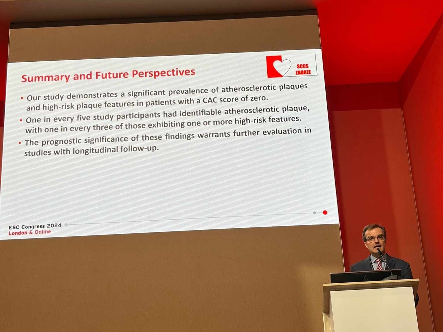
Our second study proved the limitations of this metric in accurately assessing the presence and severity of atherosclerotic burden. By employing advanced cardiovascular image analysis techniques, we were able to identify a substantial proportion of patients with a CAC score of zero who exhibited significant atherosclerotic plaques, including those with high-risk features.

The presence of high-risk plaque finally revealed
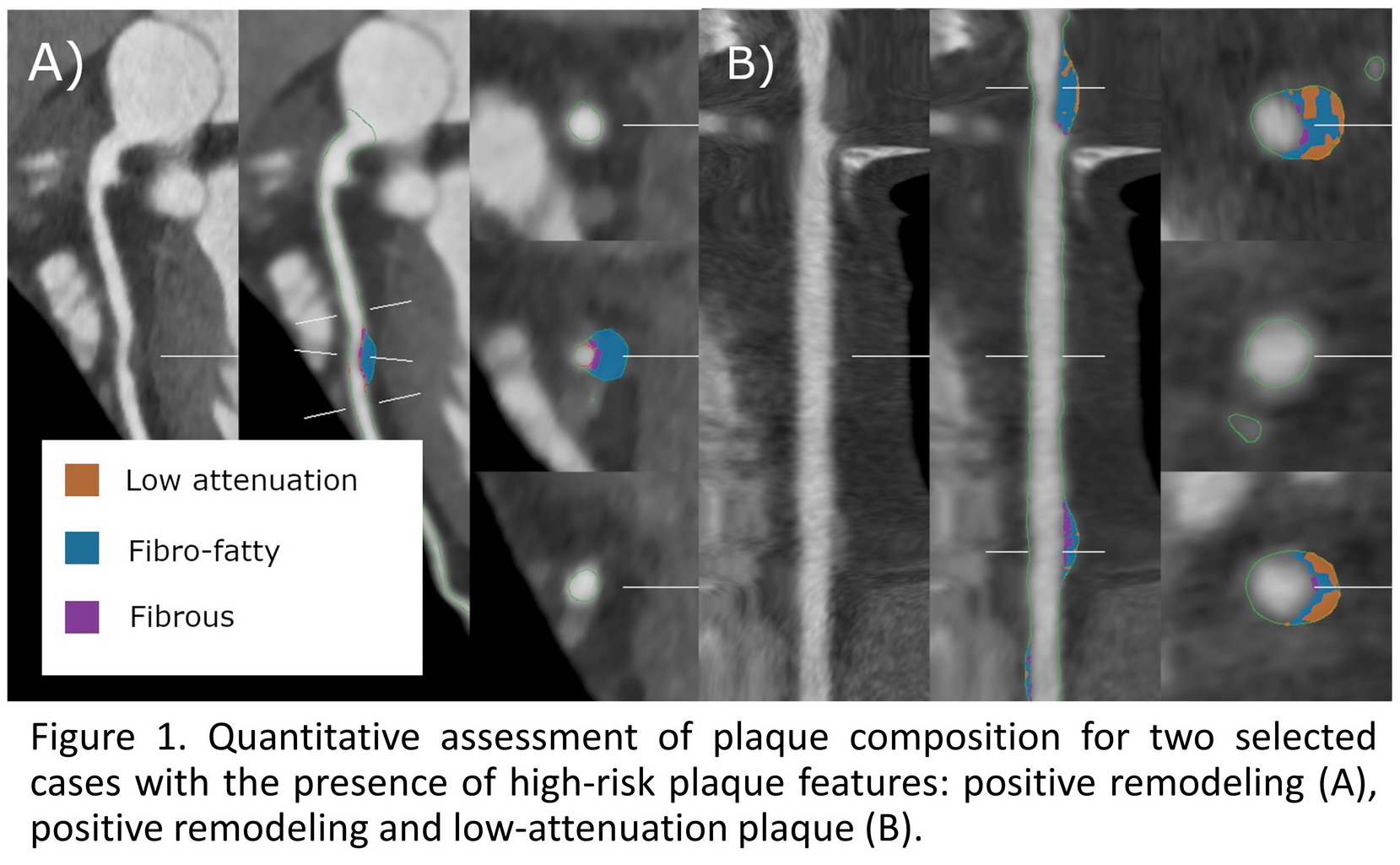
High-risk plaque features are specific characteristics of atherosclerotic plaques in coronary arteries. It indicates a heightened risk of rupture, potentially leading to a heart attack or unstable angina. More importantly, in our project, we conducted a thorough analysis of cardiac scans. We were identifying all four high-risk plaque features: positive remodeling, spotty calcification, low-attenuation plaque, and napkin ring sign. Of the 145 patients analyzed, 30 had at least one coronary artery plaque. Furthermore, in 10 of these patients, we found a plaque with at least one high-risk feature. Surprisingly, in 3 patients, we detected calcified plaque, even though the previous examination, conducted according to the Agatston criteria, showed a CAC score of 0.

AI-powered cardiovascular image analysis for a better preventive cardiology
Our findings challenge the conventional understanding of the “power of zero” and emphasize the need for more comprehensive approaches to risk assessment. While a CAC score of zero remains a valuable prognostic marker, it should not be interpreted as a definitive guarantee of cardiovascular health. A more detailed discussion of this subject can be found in our previous post titled Coronary artery calcification and… beyond calcification.
Our study presented at the ESC Congress in London underscores the importance of cardiovascular image analysis in guiding personalized risk stratification, and management strategies. It might be an extremely important tool for patients but also for the preventive cardiologists all over the world. Finally, it might be a great weapon to fight the most dangerous serial killer in the world – heart diseases.