Automating the aortic valve calcium scoring process
Aortic stenosis and aortic valve calcium scoring
Aortic stenosis stands out as the predominant primary valvular ailment necessitating surgical or transcatheter intervention in developed countries. Furthermore, this narrowing reduces the amount of blood that can flow from the heart to the body. There are two main types of aortic stenosis: the rheumatic and the calcific one, caused by a buildup of calcium on the aortic valve.
Cardiovascular imaging aids in diagnosis. Cardiac CT (CCT) is the primary test for diagnosing this condition. It is employed to quantify aortic valve calcification. This examination allows for the evaluation of aortic stenosis severity, monitoring of disease progression, and prediction of major cardiovascular events.
Where is the catch?
The diagnostic process described above seems quite straightforward, doesn’t it? However, calcium deposits often manifest in various areas of the aorta and heart. This results in misleading regions and an inaccurately computed aortic valve calcium score. Newer imaging techniques and scanners are being developed that may be able to better overcome these obstacles. However, in the meantime, we can’t stand idly by.
In one of our projects, we addressed the challenge of eliminating false-positive regions (calcifications detected outside the aortic valve) from CCT scans. We implemented a Particle Swarm Optimization (PSO) algorithm specifically designed for this purpose.
The algorithm for aortic valve calcium score
Clinicians find quantifying aortic valve calcification helpful. It aids in assessing stenosis severity where Doppler echocardiography, the primary method of assessment, does not yield conclusive results. Nevertheless, manual segmentation of aortic calcifications limits medical exam speed and accuracy. It has become an important challenge due to the growing importance of aortic valve calcium scoring in clinical practice.
Firstly, to identify potential calcifications of the aortic valve, our project utilizes anatomical information about the aorta using a deep neural network for aorta segmentation (Fig. 1). This network is then applied to a contrast CT scan co-registered to the corresponding non-contrast image. Thus, we obtained high-quality segmentation of the aortic root and aortic valve. The next step is to implement a PSO algorithm to remove calcifications that are located outside of the valve and may impact the aortic valve calcium score.

Fig. 1: A flowchart of the automated aortic valve calcium scoring.
Study’s cohort
We conducted a study on 30 pairs of contrast and non-contrast CCT scans to demonstrate the effectiveness of our end-to-end approach. We obtained the images from three different institutions that used scanners from four different manufacturers. A medical expert with 18 years of experience assessed the quality of segmentation and validated our approach. The scans come with segmentation masks for the aorta and aortic root.
The results: a precise calculation
Precise calculation of aortic valve calcification from CCT in a reproducible, user-independent, and unbiased manner holds clinical significance, facilitating improved patient selection for surgical interventions.
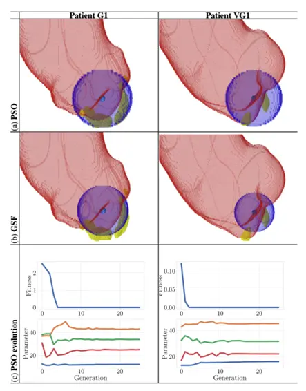
Addressing this challenge, we proposed a comprehensive end-to-end procedure for task automation. This approach incorporates a cascaded deep learning method for aorta segmentation. It is followed by thresholding the aorta’s 3D mask to identify calcification candidates. We applied Particle Swarm Optimization (PSO) to remove calcifications outside the valve (see Fig. 2). This ensured an accurate aortic valve calcium score. In our experiment, an experienced clinician rated the results for all 30 analyzed CT scans as good or very good. Additionally, this demonstrates the technique’s potential real-world usefulness.

Fig. 2: Results from (a) PSO, rated as either good (G) or very good (VG) by a reader, and the corresponding (b) GSF (with calcifications rendered in yellow).
A detailed description of the project can be found in the following paper: Jaroslaw Goslinski, Filip Malawski, Mariusz Bujny, Marcin Kostur, Karol Miszalski-Jamka, Jakub Nalepa, Deep learning meets particle swarm optimization for aortic valve calcium scoring from cardiac computed tomography, 2023 IEEE International Conference on Image Processing (ICIP), DOI: 10.1109/ICIP49359.2023.10223100.