Cardiovascular imaging and AI-enabled cardiac image processing
Benefit from state-of-the art custom software solutions and the power of AI to fight heart disease.
AI can detect hidden patterns and irregularities that may escape human notice, resulting in more accurate and timely treatments. Our diagnostic machine learning models are rooted in a deep understanding and research of the human heart and cardiovascular system.
We’ll assist you in research and software development.
Cardiovascular imaging – innovation solutions

Driving innovation across cardiology
At Graylight Imaging we bring expertise in both technology and medicine to create custom cutting-edge medical image analysis software for cardiology. We have successfully handled a number of cardiac projects and powered cardiological applications with state-of-the-art solutions.

Artificial Intelligence in cardiac
AI algorithms are a great way to improve medical decision-making and diagnostic abilities through automated cardiac image analysis. You can advance your pathway from AI-enabled image reconstruction to segmentation and precise, patient-specific measurement in various cardiac imaging modalities.
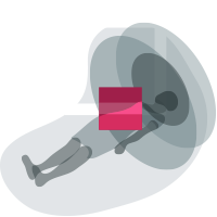
Post-processing technology for cardiac imaging
Discover advanced post-processing techniques and tools and dive into the myriad of data embedded in cardiovascular images. AI-enabled cardiac image processing can transform patient care at every stage of the imaging chain. AI methods offer the extraction of new, clinically relevant information for patient and risk assessment.
Development of cardiac imaging solutions – selected work
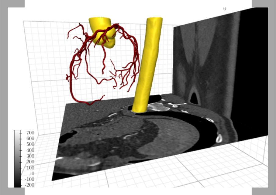

AI algorithms for accurate segmentation of coronary vessels for cardiovascular imaging
Accurate segmentation of cardiovascular area allows creation of a personalized treatment plan, with unique anatomy and tumor location. It enables more precise and effective treatment while minimizing damage to surrounding healthy tissue.
Creating technology for automatic coronary artery segmentation was one of the biggest challenges. This was due to several factors: complicated shape of coronary arteries and patient variability in coronary arteries, particularly in anomalies.
Despite difficulty, we have developed a technique for creating AI algorithms for precise coronary artery segmentation.
Our models for automatic segmentation of coronary arteries reconstruct the coronary artery tree with an accuracy of more than 90% (Sørensen-Dice coefficient of about 0.9).
Our algorithms have also found application in cardiology. In the case of cardiovascular lesions, accurate image segmentation enables more effective diagnosis and treatment planning.

Analysis solutions for cardiovascular imaging and coronary artery calcification
The amount of calcified (hard) plaque in your heart vessels can be determined quickly, conveniently, and noninvasively with a cardiac calcium score computed tomography (CT), also known as a coronary calcium scan.
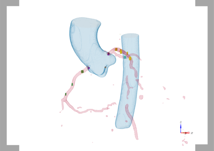
In one of our projects, we faced the challenge of developing an algorithm that not only segments the plaques and calculates the Agatson score, but also assigns calcified plaques to the appropriate part of the cardiovascular tree.
End-to-end automation of this process was key, as was precision – the goal was to be accurate to within 1 voxel.
The result is presented in the graphic (different colors of plaques are assigned to the corresponding part of the vascular tree).
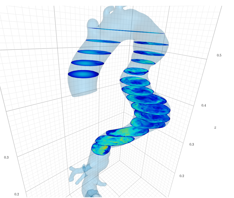
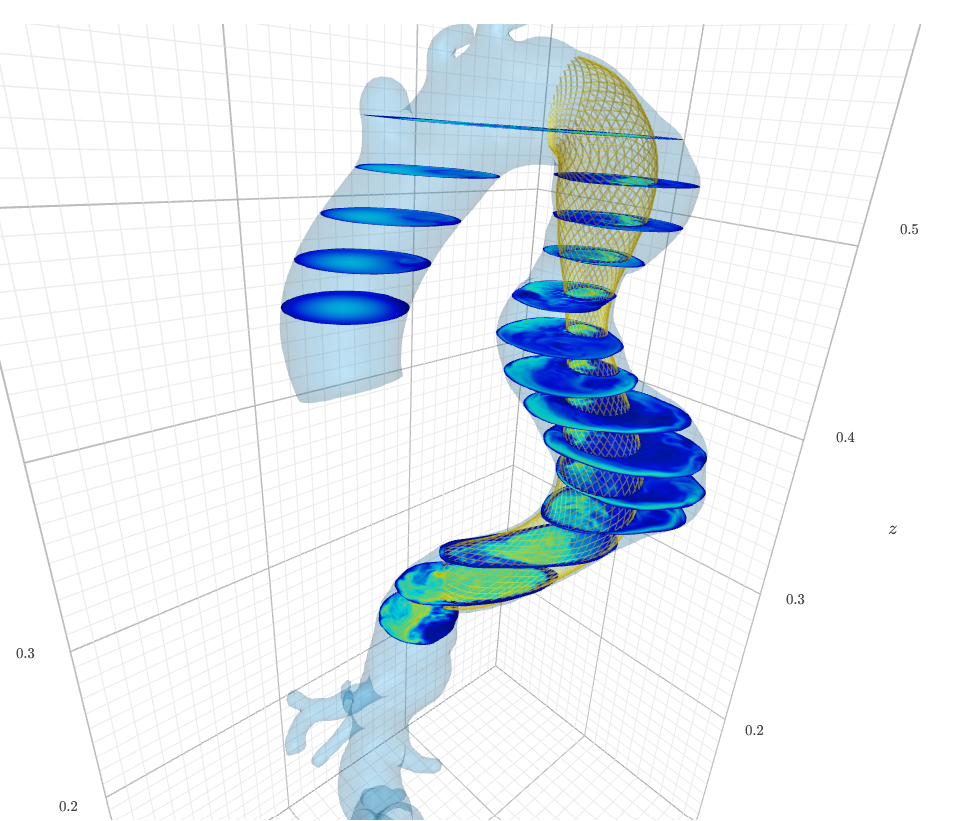


Computational fluid simulation (CFD) of flow in aortic aneurism before and after stenting
In interventional cardiology, artificial intelligence and biofluid simulation methods have shown the potential to provide data interpretation and automated analysis, as you may see in the graphic.
Tools for non-invasive assessment of cardiovascular conditions may benefit from the newest technology and cardiovascular imaging analysis.
Advanced techniques and analysis solutions for cardiovascular imaging might be applied for interventional cardiology purposes as well.
During the project, based on the CT scans we provided a fully automated, patient-specific blood flow simulation before and after the stenting procedure.

AI assessment in cardio imaging using centerline-based coronary vessels segmentation
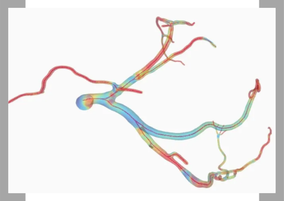
We have developed a comprehensive deep learning-based pipeline for automated analysis of coronary computed tomography angiography using computational fluid dynamics (CFD).
We use centerline-based segmentation rather than manual segmentation. As a result, we get blood flow metrics that are in great agreement with created for ground-truth delineation.
A centerline is a line that connects two points and runs through the middle of the model. They shed a lot of light on its topology. As a result, can determine the diameter of the vessel at each point along the centerline.
We can claim that such a solution performs better than cutting-edge nnU-Nets.
Proven experience in the cardiovascular imaging field
CUTTING-EDGE TECHNOLOGIES FOR CARDIAC IMAGING AND ANALYSIS
At Graylight Imaging, we thrive on innovation and we’re ready to team up with you to fight the leading killer in the modern era: heart disease. We can integrate clinical and technological knowledge to boost your solution by leveraging multiple advanced image processing methods and techniques as well as analysis solutions for cardiovascular imaging. Check one of our latest papers.


