Automatic brain tumor segmentation with subregions
Our models automatically perform brain tumor segmentation and segment 3 tumor subregions (edema, enhancing tumor and necrosis) and provide volume and RANO (Response assessment in neuro-oncology) measurements. So, it can be a useful tool for monitoring patients in clinical studies.
Moreover, we are able to develop custom automated and semi-automated algorithms that analyze the brain and brain lesions, according to your need. As a result, the following example illustrates how our algorithms have been used to assess brain tumor segmentation, treatment and progression.
Our work: AI algorithm for brain tumor segmentation
In this paragraph, we would like to focus on the following example, which demonstrates the limitations of RANO measurements. Firstly, when compared with full volumetric measurement of a segmented image based on the Brain-Tumor-Progression dataset, we show how our models can be used to assess brain tumor treatment and progression. Secondly, in the two examples below, we visualize how our models can be used for fully-automated tumor monitoring in longitudinal studies and what information can be extracted.
In addition, for each patient, we present the most representative slice of the tumor (usually the one selected for RANO calculation) and compare the results of our model in two-time points. Our models require MRI studies with 4 sequences (T1-pre, T1-post, T2, FLAIR) but for visualization purposes, we present only T1-post and FLAIR series. The segmentations presented in the examples were also approved by our cooperating senior radiologist.
Visualization: Brain tumor segmentation: regression
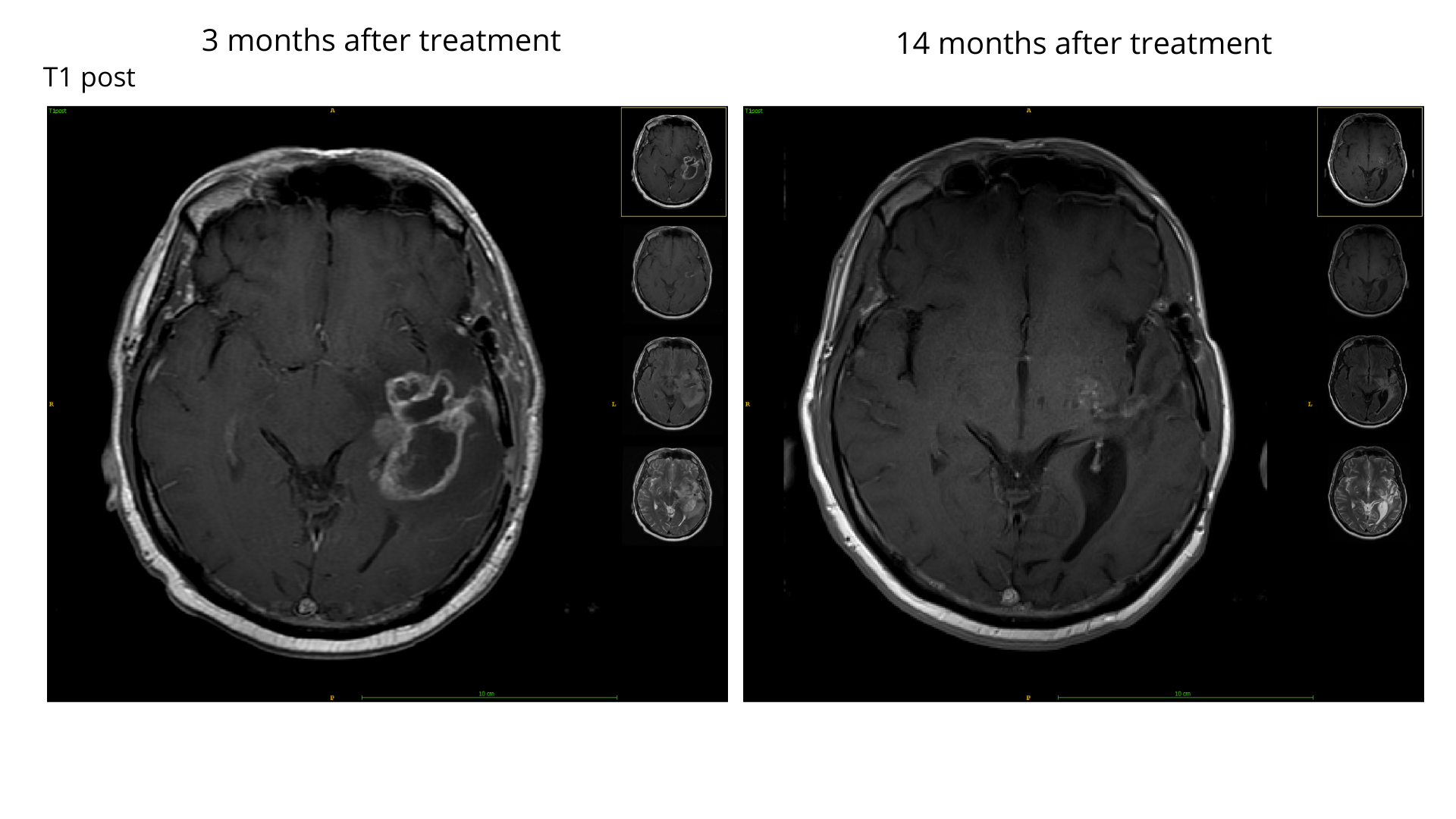
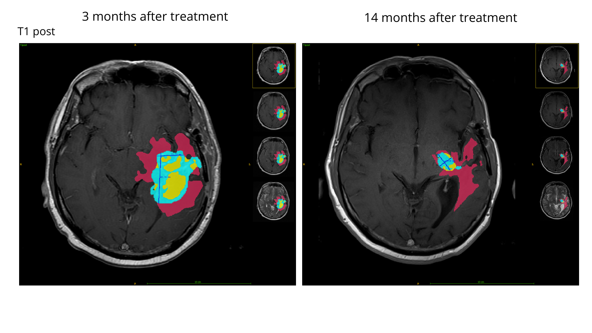
The patient responded well to the treatment, and we observed a strong enhancing tumor (ET) regression for this patient a year after the treatment. Automatic RANO measurement (marked with blue line segments in both scans) and ET volume both significantly decreased (-78% and -93% respectively). In summary, the region of edema became much smaller, and necrosis was nearly absent.


Edema: 97 cm3
Enhancing (ET): 28 cm3
Necrosis: 19 cm3
Tumor regression
RANO -78%
ET volume -93%
Edema: 39 cm
Enhancing (ET): 2 cm3
Necrosis: 1 cm3
Brain tumor segmentation: progression
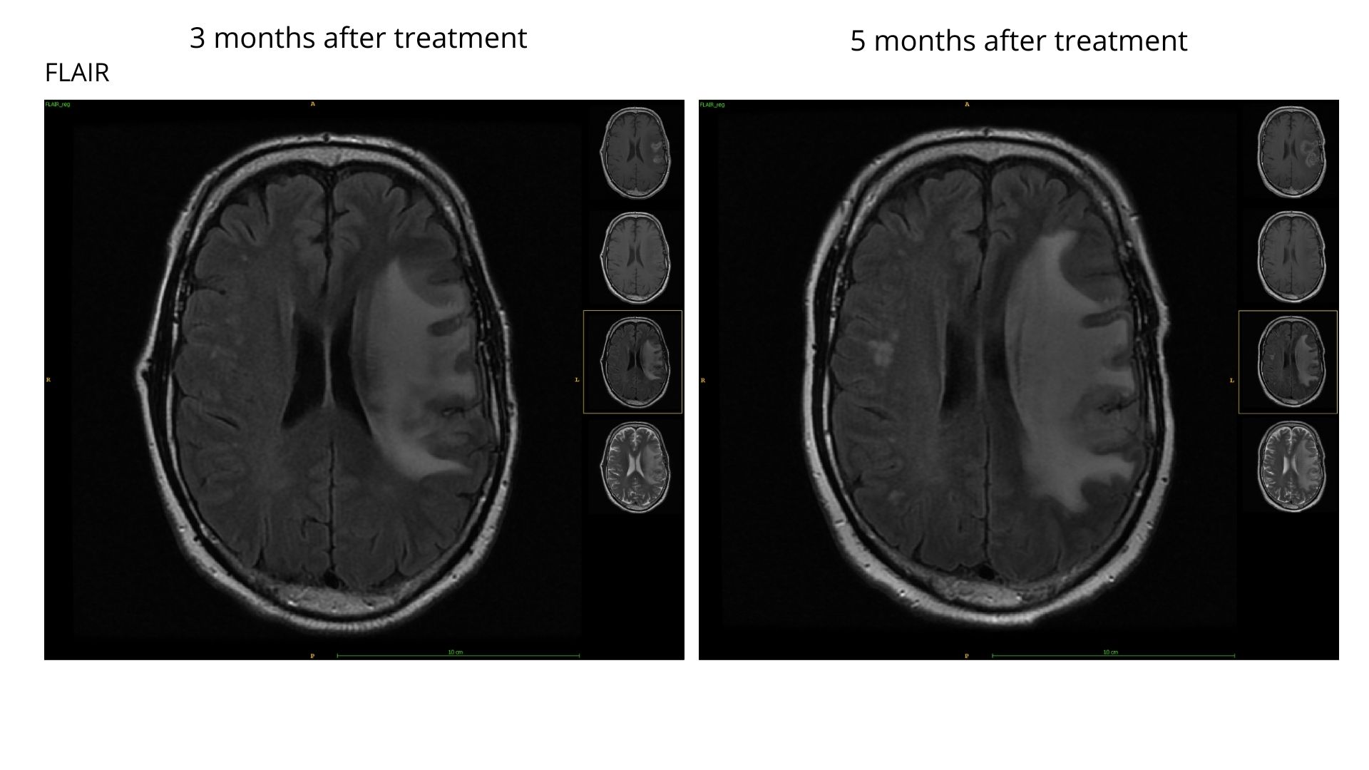
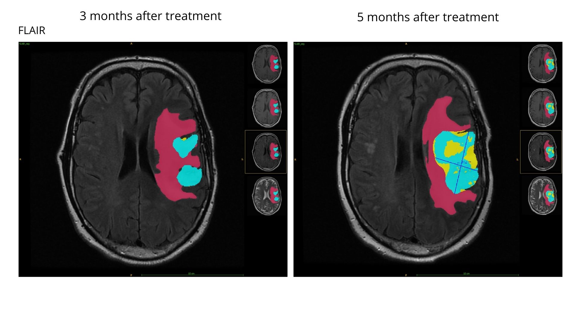
In this case, our model showed a doubled tumor burden in just 2 months between scans for this patient. Two separate enhancing tumor regions have grown and merged into one big lesion. It is reflected in RANO measurement marked in the later scan (RANO for the earlier scan was measured on a different slice). In sum, all the subregions have expanded what confirms the rapid tumor progression.


Edema: 82 cm3
Enhancing (ET): 20 cm3
Necrosis: 2 cm3
Tumor progression
RANO +100%
ET volume +85%
Edema: 136 cm3
Enhancing (ET): 36 cm3
Necrosis: 20 cm3
Would you like to learn more about our competencies? Visit the website with the publication:
Deep learning automates bidimensional and volumetric tumor burden measurement from MRI in pre- and post-operative glioblastoma patients