Cardiac image processing – novelties and more
Cardiac image processing is a crucial field in AI-powered medical imaging. It plays a pivotal role in personalized medicine. Unsurprisingly, this year’s edition of The European Society of Cardiology Congress, which spotlights ‘Personalizing Cardiovascular Care’, invites to several interesting sessions dedicated to advanced medical image analysis. It would be also a great chance to get familiar with some of our projects. Karol Miszalski-Jamka, MD, PhD will deliver two presentations:
- AI-based algorithm for assessing coronary artery calcium score on contrast-enhanced cardiac computed tomography scans
- Prevalence of high-risk plaque features on cardiac computed tomography scans in patients with a coronary artery calcium score of zero
Coronary artery disease. The importance of early detection
Coronary artery disease (CAD) is a leading cause of death worldwide, responsible for approximately 610,000 deaths annually in the United States alone. In fact, it accounts for about one in four deaths in the country. Moreover, the healthcare costs associated with CAD in the US are estimated to exceed 200 billion dollars annually. [1]
Despite its severity, CAD is curable, especially when diagnosed early. Therefore, when emphasizing the importance of early intervention, one cannot omit the role of cardiac image processing and advanced analysis.

Cardiac image processing to predict CAD events
Early detection of individuals who are at high risk for heart disease but have no symptoms is crucial for maximizing the effectiveness of preventive medications like statins. Current CAD assessment with Computed Tomography encompasses two examinations: Coronary Artery Calcium Score and Coronary Artery CT Angiography. Angio-CT allows doctors to evaluate narrowings and obstructions in the coronary arteries. The coronary artery calcium (CAC) score is a measure of plaque buildup in the coronary arteries. It can accurately predict future adverse heart events.
This means that a patient undergoes CT examination twice and is exposed to ionizing radiation twice. A relevant query is whether two distinct CT scans are truly needed to assess both coronary arteries and calcified plaques… Thus, can we reduce this unnecessary patients’ radiation exposure?
AI in cardiac image processing. Calcium score case
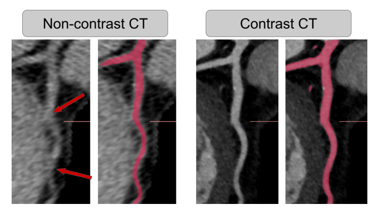
Of course, the struggle behind double examinations is unfortunately quite simple. While a contrast agent helps visualize coronary arteries, it can sometimes mask calcified plaques. However, AI-powered cardiac image processing opens many doors that have been closed before.
In one of our projects, we exploited the possibilities offered by AI algorithms and decided to check if coronary artery calcium score can be obtained based solely on contrast-enhanced CT scans. Karol Miszalski-Jamka MD, PhD will present this AI-based algorithm and its results during the poster session on 2nd September.

Figure 1: The challenge of segmenting the coronary vessel from non-contrast CT, image source: Bujny, K. Jesionek, J. Nalepa, K. Miszalski-Jamka, K. Widawka-Żak, S. Wolny, M. Kostur: Coronary artery segmentation in non-contrast calcium scoring CT images using deep learning.
Patients with a coronary artery calcium score of zero
It is often believed that a CAC score of zero, often referred to as “power of zero,” is linked to a very low risk of serious heart problems. Particularly in the short and medium term. However, the MESA Study showed that even individuals with a CAC score of zero still experience a significant number of major heart events. The number of those patients accounts for about one-fourth to one-third of the total.
It is known that CAC score of zero does not definitively exclude the presence of non-calcified atherosclerotic plaques or narrowings in the coronary arteries. We aimed to understand the composition of atherosclerotic plaques and how often dangerous plaque characteristics (so-called high-risk plaque features) are seen on contrast-enhanced CT scans in people with a CAC score of zero. Despite being a very challenging project, the team was driven by the significance of the potential findings.
As of September 1st, you will have the opportunity to review the outcomes of our work [2]. Don’t miss the session ‘Novel assessment of cardiovascular risk using computed tomography’.
The London’s journey to the heart
The European Society of Cardiology Congress starts on August 30th in London. It is a unique opportunity to connect with the cardiovascular community, explore cutting-edge research, and gain insights into personalized patient care. And to know more about our work in cardiac image processing field. See you in London!
References:
[1] Brown JC, Gerhardt TE, Kwon E. Risk Factors for Coronary Artery Disease. 2023 Jan 23. In: StatPearls [Internet]. Treasure Island (FL): StatPearls Publishing; 2024 Jan–. PMID: 32119297.
[2] https://esc365.escardio.org/ESC-Congress/speakers/206323