Artificial intelligence in medical imaging: from algorithm to diagnosis
Once a concept confined to computer science labs and speculative fiction, artificial intelligence is now transforming medicine, especially radiology and medical imaging. But this transformation has been anything but overnight. From theoretical beginnings in the 1940s to today’s deep learning-powered systems capable of interpreting complex scans, AI’s journey and medical algorithms development into medicine is a story of persistence, innovation, and scientific breakthroughs.
This article traces that journey – from the earliest models and expert systems to the rise of neural networks, deep learning, and the challenges that lie ahead.
The post is based on a scientific paper: The evolution of artificial intelligence in medical imaging: from computer science to machine and deep learning, 2024.
The foundations of artificial intelligence in medical imaging: from Turing to the first expert systems
The roots of AI trace back to a simple question posed by Alan Turing in 1950: “Can a machine think?” His famous Turing test introduced the idea that computers could imitate human reasoning. This sparked decades of exploration into symbolic reasoning and artificial neural networks.
Symbolic AI, which relied on logic-based IF-THEN rules, dominated early efforts. It worked well in structured environments like games, but struggled in dynamic settings like healthcare. In contrast, machine learning introduced a data-driven approach, allowing systems to learn from patterns and experience, better suited to complex medical information.
By the 1960s, researchers like Frank Rosenblatt developed the perceptron, an early neural network model capable of basic image classification, seen by many as the first step toward AI in image analysis. Still, early models lacked the sophistication and scale required for practical use.
In the 1970s and 1980s, expert systems emerged as rule-based programs designed to support clinical decisions. While groundbreaking at the time, they had major limitations: they couldn’t adapt to new data, and they struggled with tasks requiring flexibility, like interpreting variable medical images.

The “AI Winter” and the path to deep learning
Enthusiasm for neural networks declined in the late 1960s after it was shown that single-layer models couldn’t solve complex problems, a period known as the AI winter. For years, funding and interest dwindled.
That began to change in the 1990s. New algorithms, along with the spread of personal computers, helped AI regain momentum. These tools reintroduced AI to medical researchers, but the real breakthrough came with the emergence of deep learning.
Neural networks reborn: AI meets medical imaging
AI’s entry into radiology began modestly. In the 1980s, researchers used simple neural networks to assist with interpreting radiological data, including mammograms. These early tools needed manually crafted features and offered limited precision, but they planted the seeds of transformation.
By the late 1990s and early 2000s, Computer-Aided Diagnosis (CAD) systems became more common in clinical settings. These programs highlighted suspicious areas in X-rays or MRIs, acting as a second set of eyes for radiologists. However, they often produced too many false positives, lacked transparency, and couldn’t learn or improve over time.
The real revolution began with the rise of deep learning, particularly convolutional neural networks (CNNs). CNNs excel at image analysis, learning to identify features directly from raw data without human-defined rules. Combined with advances in GPU computing, open-source libraries (like TensorFlow and PyTorch), and access to large image datasets, deep learning opened the door to high-performance AI in medical imaging.
Today’s AI models can:
- Detect cancerous changes in images with accuracy comparable to human experts,
- Classify tumors and tissue types,
- Segment anatomical structures in CT and MRI scans,
- Track disease progression,
- Evaluate treatment response over time.
From support tool to diagnostic partner
Recent studies show that in some scenarios, such as screening mammography, AI can equal or outperform two human radiologists working together. This shifts AI from a passive assistant to an active diagnostic partner.
Beyond imaging, AI technologies like natural language processing (NLP), recurrent neural networks, and generative models allow for automatic data labeling, medical report analysis, and integration into clinical workflows. With affordable computing power and open-source tools, even smaller research centers and hospitals can deploy custom AI solutions.
Open challenges: what’s holding AI back?
Despite the progress, significant challenges remain before AI can be fully integrated into everyday clinical use:
a. Clinical validation
Many AI systems perform well in research but haven’t been tested in real-world, clinical environments. Prospective trials in mammography, are crucial steps forward, but more multi-center, randomized studies are needed.
b. The black box problem
AI models often lack transparency. Clinicians may hesitate to trust decisions they can’t interpret. This has driven interest in explainable AI – methods like heatmaps and saliency maps that highlight what the algorithm focused on when making a decision.
c. Ethics and regulation
AI systems must be fair, unbiased, and accountable. Unequal training data can introduce bias. Clear legal frameworks are also needed. Initiatives like the EU’s Artificial Intelligence Act aim to standardize ethical use and ensure safety.
 In radiology, deep learning models can now match expert performance in many diagnostic tasks, offering faster, more consistent, and scalable analysis."" title="AI in medical imaging from algorithm to diagnosis" srcset="https://graylight-imaging.com/wp-content/uploads/2025/08/AI-in-medical-imaging-from-algorithm-to-diagnosis.png 768w, https://graylight-imaging.com/wp-content/uploads/2025/08/AI-in-medical-imaging-from-algorithm-to-diagnosis-300x169.png 300w, https://graylight-imaging.com/wp-content/uploads/2025/08/AI-in-medical-imaging-from-algorithm-to-diagnosis-120x68.png 120w" sizes="(max-width: 768px) 100vw, 768px" class="wp-image-284106 webpexpress-processed">
The future: toward multimodal, personalised medicine
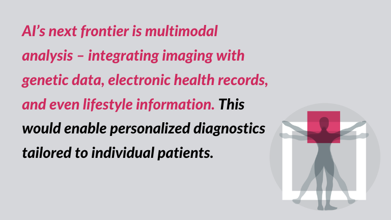
AI’s next frontier is multimodal analysis – integrating imaging with genetic data, electronic health records, and even lifestyle information. This would enable personalized diagnostics tailored to individual patients.
New projects are pushing boundaries by developing systems that can learn across various data types simultaneously, mimicking the human ability to synthesize complex inputs.
Meanwhile, traditional machine learning (like decision trees) still has a place, especially where transparency, low cost, and simplicity are essential.
Conclusion: AI in medical imaging – a transformative force
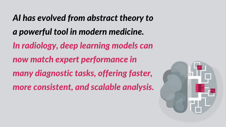
AI has evolved from abstract theory to a powerful tool in modern medicine. In radiology, deep learning models can now match expert performance in many diagnostic tasks, offering faster, more consistent, and scalable analysis.
Yet for AI to reach its full potential, it must be clinically validated, transparent, ethically sound, and regulated. Progress is promising, but continued collaboration between doctors, scientists, engineers, and policymakers is essential.
The future of AI in medical imaging isn’t just coming – it’s already here. And its potential to reshape diagnostics, improve outcomes, and personalize care is only just beginning to be realized.
Resourcres:
Avanzo M., Stancanello J., Pirrone G., Drigo A., Retico A., The evolution of artificial intelligence in medical imaging: from computer science to machine and deep learning, Cancers, 2024, https://www.mdpi.com/2072-6694/16/21/3702.