Multi-class brain tumor segmentation and volumetric measurements
The text is based on materials by Bartosz Machura
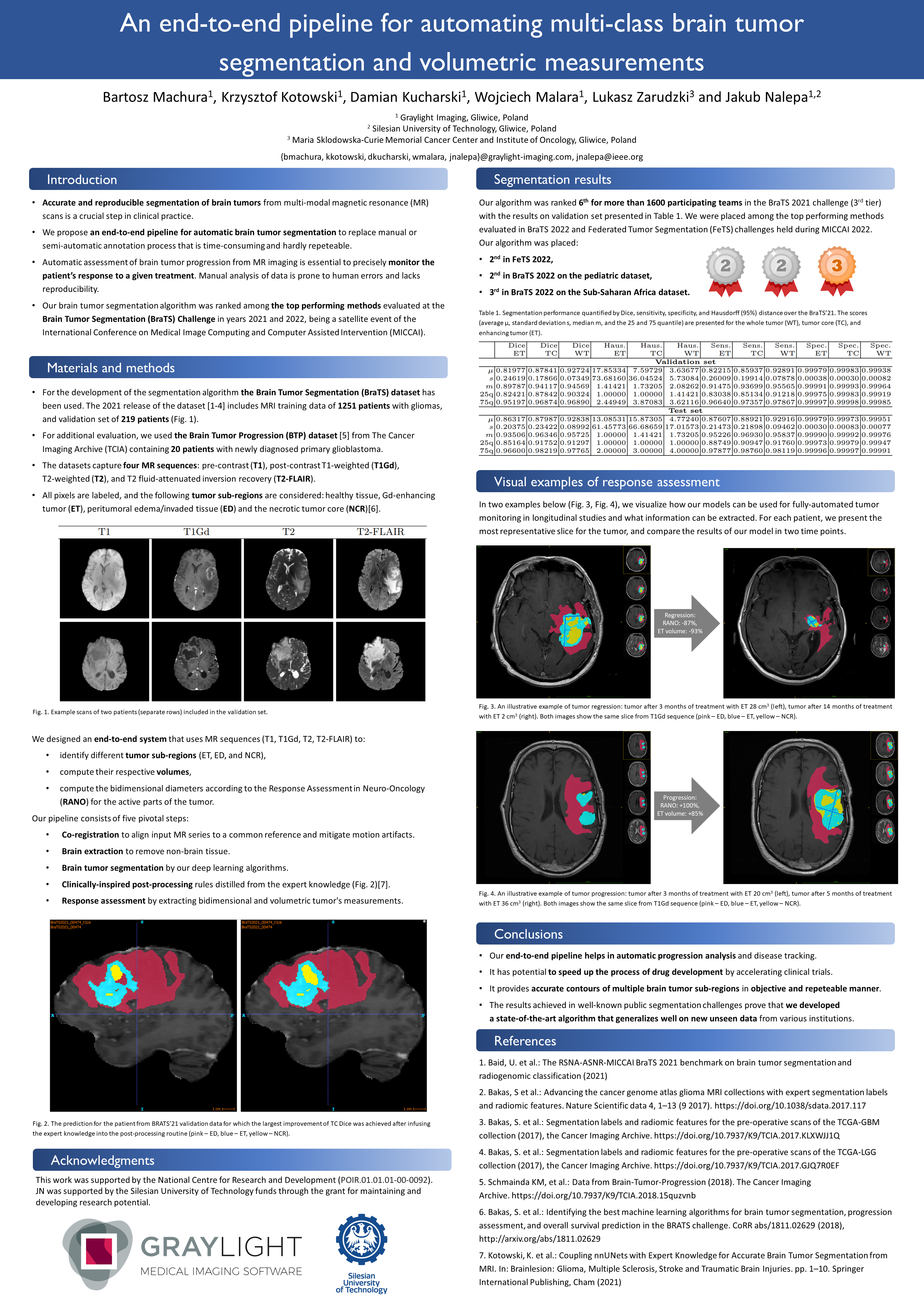
An end-to-end pipeline for automating multi-class brain tumor segmentation and volumetric AI measurements. This is the title of poster which was displayed by Bartosz Machura during the BioTechX congress. This largest European conference on diagnostics, precision medicine, and digital revolution in drug development was organized last week in Basel.
Multi-class brain tumor segmentation – our approach
Tumor burden assessment by magnetic resonance imaging (MRI) is crucial to the evaluation of glioblastoma treatment response. This assessment is complex to perform and associated with high variability due to the heterogeneity and complexity of the disease.
The introduction of a new drug requires laborious and strenuous testing in clinical settings. During clinical study experts analyze lesions from multiple visits to track and assess disease progression. Radiology experts usually contour lesions’ sub-regions manually or semi-automatically. This requires multiple radiologists. It is a time-consuming as well as hardly reproducible process.
Our deep learning-powered solution tackles this issue by offering full reproducibility of an AI tool for delineating and analyzing the brain tumor lesions which also accelerates the analysis process.
“It is worth mentioning that we established the state of the art in multi-class segmentation of brain tumors – our solution was among the top performing methods evaluated in this year’s Brain Tumor Segmentation Challenge (BraTS) and Federated Tumor Segmentation Challenge (FeTS) held during the Medical Image Computing and Computer Assisted Intervention conference (MICCAI).”
Brain tumor segmentation and volumetry
Algorithms can analyze large amounts of data to segment brain tumors more efficiently and with a higher consistency, which offer an inter-observer variability reduction. This not only saves time for medical professionals but also improves the accuracy of patient response assessment. Furthermore, the algorithm we introduced can automatically calculate tumor volume. This widely meaningful factor in monitoring tumor response has been offically introduced as optional by RANO criteria update.
AI-powered brain tumor analysis: our poster
A multi-class segmentation algorithms are similar to super-tools that can divide an image into specific categories, and then: handle a bunch of different ones at the same time. This type of algorithm is highly useful when you have to deal with images with a variety of things to identify and categorize at the same time. This is exactly why our team developed a solution based on this architecture. Below you can find the mentioned poster, presenting our multi-class brain tumor segmentation algorithm.

Read the previous post about our participation at BioTechX congress: BioTechX – our brain tumor segmentation algorithm