FEATURES EXTRACTED FROM MEDICAL IMAGES
Radiomics in medical imaging analysis
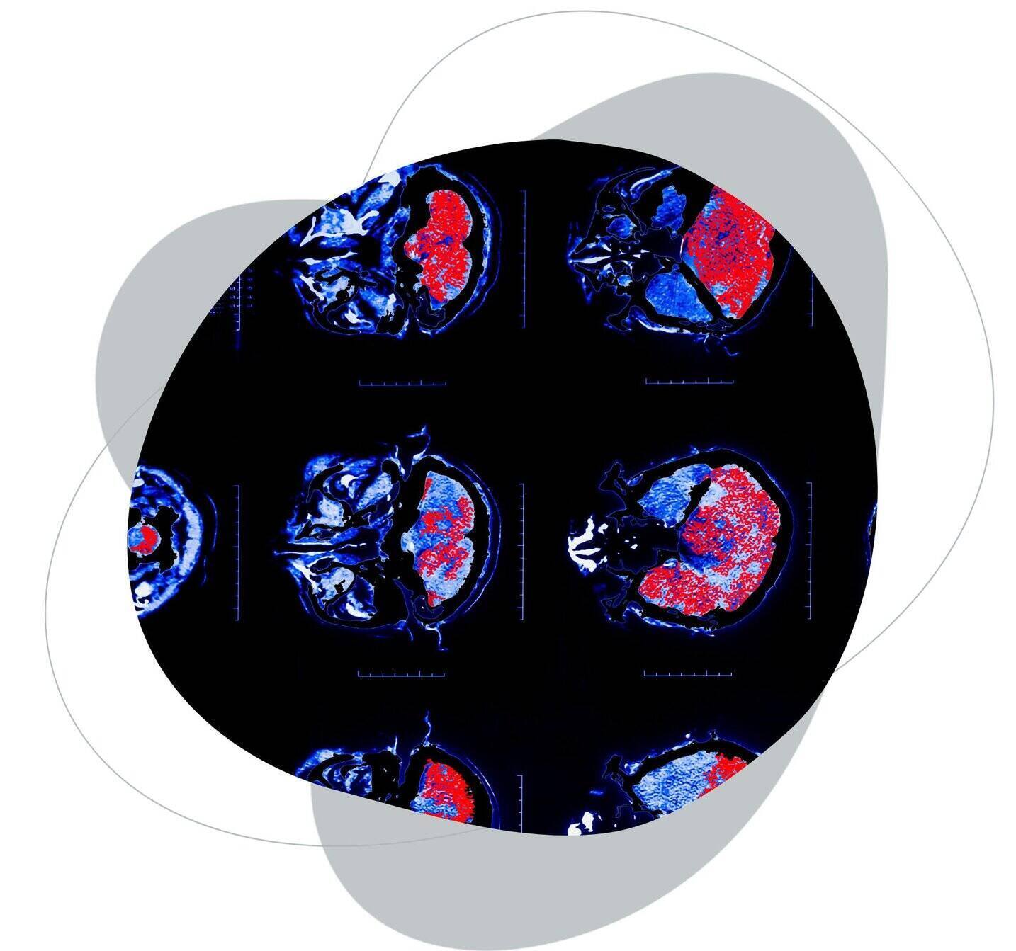
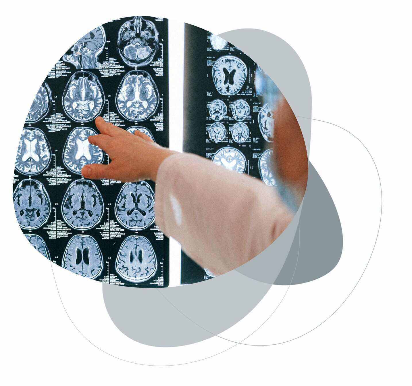
Radiomics is a method that extracts numerous quantitative features
from medical images that are unrecognizable to the naked eye.
This approach enables the creation of new imaging biomarkers
and access to additional information at a microstructural level.
Gain the advantage using Radiomics
New imaging biomarkers
Radiomics provides new imaging biomarkers for very precise and detailed exploration of pathologies. Additionally, it minimizes interobserver variability by leveraging AI-enabled segmentation and analysis.
Exposing data and patterns
This method expounds medical images to numbers which helps clinicians to see the lesions in multiple dimensions and compound them with structural and textural characteristics. It enables more accurate treatment planning by exposing data and patterns within a lesion that were hidden beforehand as well.
Better patient selection
By leveraging Radiomics you can better select and stratify patients at the very early stage in drug development trajectory. The combination of imaging and phenotypic data may reveal the key to facilitating better clinical decision-making and treatment.
Early indicators of treatment effectiveness
Data derived from images combined with texture analysis fuels diagnosis, therapy and prognosis. Radiomics provides early indicators of treatment effectiveness, which makes it a great aid in patient response assessment and clinical trials.
Custom radiomics project development

Image data acquisition and reconstruction. It is important to homogenize data during the acquisition or pre-processing step. Data preparation should be not only pixel or voxel oriented, but we also need to check that the data is unbiased to avoid modelling problems.

Segmentation – we develop precise and automated segmentation algorithms. We fully understand that this step is the most critical component of radiomics – the following feature data and quantification are generated from the segmented ROI (region of interest).

Features extraction & selection. In every project, we follow the criteria of the Image Biomarker Standards Initiative. By working closely with clinicians, we can also create handcrafted features. Selected biomarkers are analyzed by tailored algorithms.

Data analysis & interpretation. In this step, we build a model. Modelling allows us to respond to the asked question – for example to determine which patients are likely to benefit from therapy or to predict the risk of developing recurrence of a disease.
Why develop a radiomics project with Graylight Imaging?
- We have experience in the extraction, selection, and grouping of radiomic features and data analysis in a radiomic project. Thanks to prepared functions, we create a model answering the question asked.
- We create automatic, advanced, and accurate machine learning algorithms – as a basic element of the radiomic project. According to your current need, we are able to design an algorithm for your specific needs.
- We provide specialized algorithms for data characterization that analyse medical images using particular biomarkers. With the help of the algorithms we’ve created, we can extract a plethora of characteristics from medical photos.

Learn more about our competences
Contact us
Let’s talk
We’d love to discuss your needs, ideas, or challenges.


