Segmentation of the thoracic aorta in contrast enhanced CT images
The challenge: an algorithm for thoracic aorta segmentation
In one of our projects we faced the challenge of the development of AI algorithm for the segmentation of thoracic aorta in contrast enhanced cardiac computed tomography (CT) images.
Contrast-enhanced CT images are the preferred method for the assessment of thoracic aorta including visualization of pathologies (e.g. aortic aneurysm*) and measurement of diameters at specific levels. Moreover, accurate segmentation and visualization of aortic root is of special interest in cardiac CT imaging. Aortic root is the proximal portion of ascending aorta beginning at the level of aortic annulus and extending to sinotubular junction. Between each commissure of the aortic valve there are usually three aortic sinuses.
Why is this a difficult task?
Examining the shape (morphology) of the aorta and identifying any abnormalities (pathologies) from CT scans is crucial for diagnosing heart problems and assessing a patient’s risk. However, doing this manually on CT scans with low radiation and no contrast dye is very slow and difficult.
Automatic detection of the aortic root is an important element for further AI data processing. The difficulty in automatically segmenting the aortic root is twofold. Firstly, the aortic valve is poorly visible and classical methods of segmentation based on thresholding usually fail. Secondly, the valve can be opened and then the contrast connects directly to the left ventricle. In both cases, radiologists and cardiologists can rely on the general anatomical features of aortic sinuses/aortic annulus to extrapolate the location of a poorly visible aortic valve. The challenge is to create an algorithm that incorporates this intuition and correctly segments the aortic root in both cases.
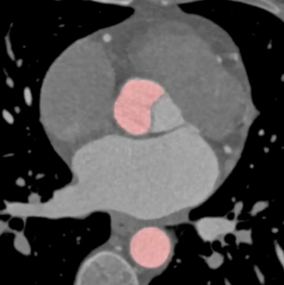
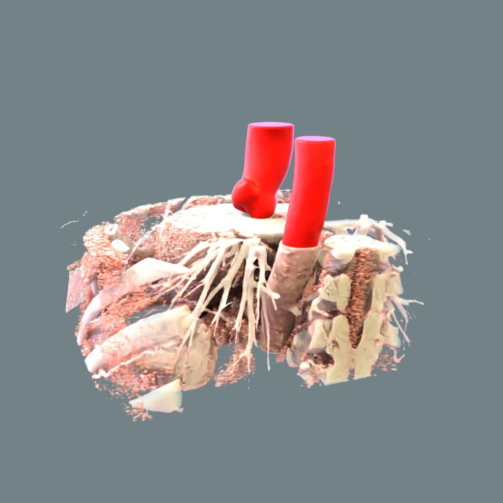
ML generated segmentation of thoracic aorta. Note the accurate segmentation of aortic cusps.
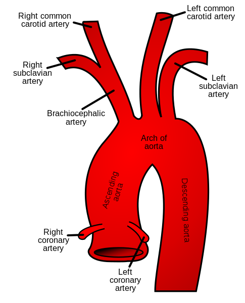
Methods we used
Our team consisted of experts from various fields – software engineers, researchers, clinicians, and physicists faced the need to develop the custom algorithm. Data for the algorithm were prepared using classic image segmentation techniques such as Fast Marching Methods. Additionally, we used the results of the prepared ground truth to create an algorithm based on standard machine learning solutions.
The segmentation of the thoracic aorta algorithm performance
We always evaluate the accuracy of an artificial intelligence-based algorithm. It is an important step, especially in feasibility projects.
Here we developed an AI algorithm for the segmentation of the thoracic aorta on contrast-enhanced cardiac CT images. The average Sørensen-Dice [1] coefficient of the model was above 0.98. The model is robust across images of different quality and produces meaningful results even in the case of open valve.
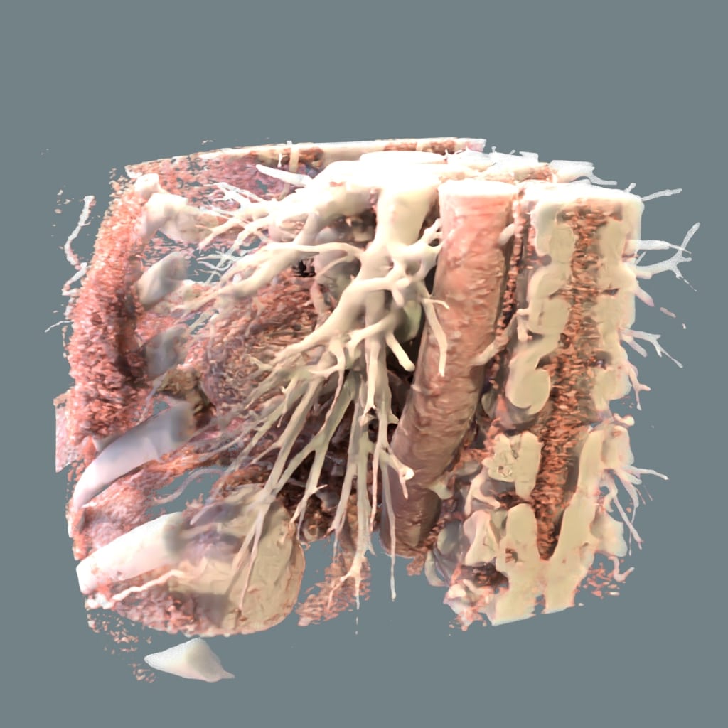
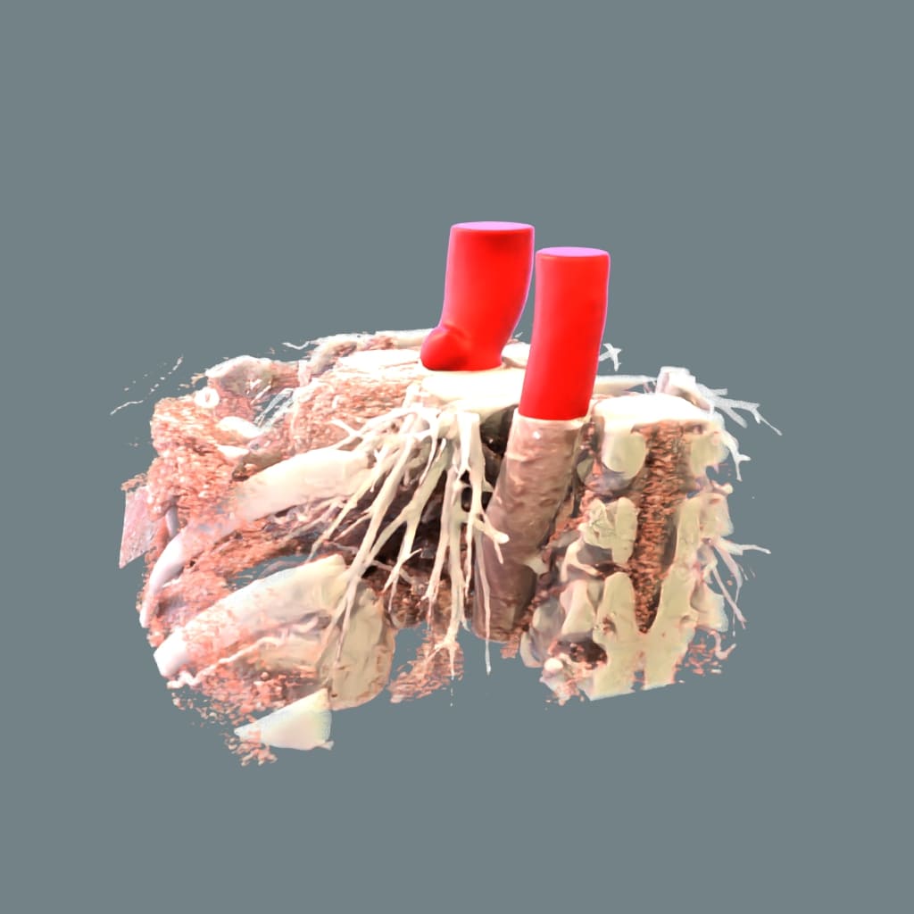
What is a thoracic aortic aneurysm?
*An aneurysm develops when a weak spot in an artery wall stretches and balloons outward. The aorta, the body’s biggest artery delivering oxygen-rich blood from the heart, is particularly vulnerable. Aortic aneurysms can occur in two main places: the chest (thoracic) or the abdomen (abdominal).
Several things can raise a patient’s risk of developing a thoracic aortic aneurysm. The most common causes include:
- ageing,
- high blood pressure,
- cholesterol buildup (atherosclerosis),
- aortic dissection
References:
[1] https://en.wikipedia.org/wiki/Dice-S%C3%B8rensen_coefficient