Liver and liver tumor segmentation and analysis algorithms

We are able to develop custom automated and semi-automated algorithms to liver and liver tumor segmentation and analysis, according to your needs.
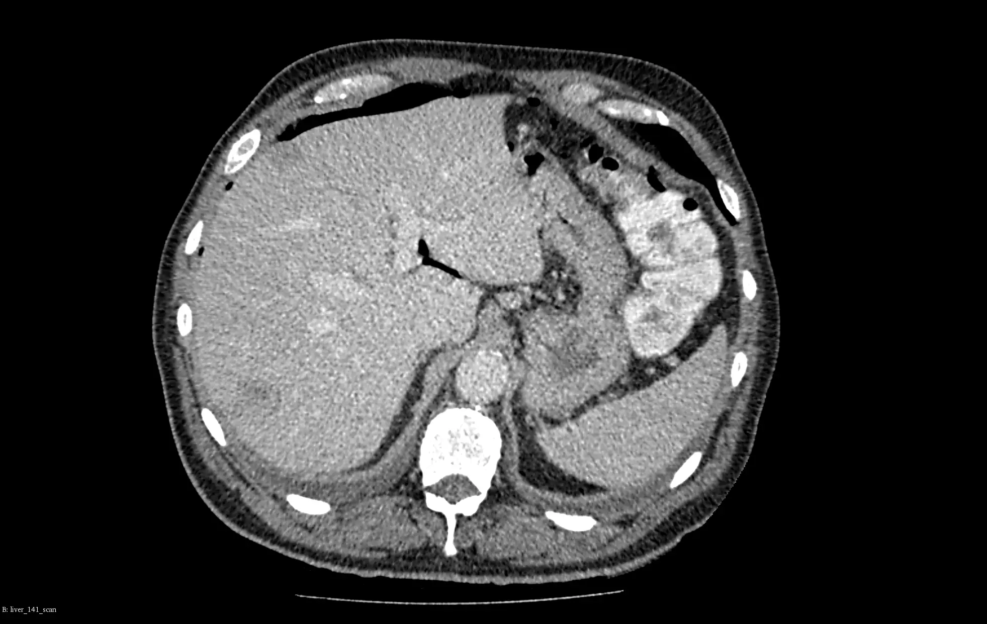
We present our models for liver and liver tumors segmentation (including primary tumors, secondary tumors and metastases) based on portal venous CT scans.
OUR WORK
AI algorithm for liver tumor segmentation
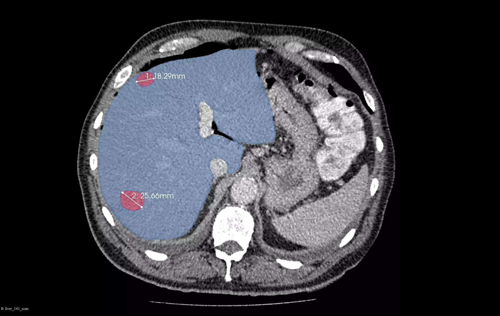
In the example below we vizualize how our models work for a patient from LiTS2017 test set (not used for training). Our algorithms automatically perform volume and RECIST measurements of the tumors with an option to extend it to mRECIST if proper training data are available. It can be a useful tool for monitoring patients in clinical studies.
Our liver segmentation model was trained on a dataset of more than 500 patients. Its results were used to extract the region of interest for tumors segmentation which was trained on more than 400 patients. Our tumor segmentation model is in the current top best solutions in terms of average Dice (0.77) on LiTS2017 Challenge Open Leaderboard. The liver segmentation model provides very accurate results with an average Dice of 0.954 on LiTS2017 test data.
VISUALIZATION
Liver and the liver tumor segmentation
- The liver segmentation is marked in blue
- The tumor segmentation is marked in red.
- The RECIST measurements are marked in white.
- The models identified 4 measurable tumors with longest diameters of 26 mm, 18 mm, 16 mm, and 10 mm, and with corresponding volumes of 9 ml, 2.3 ml, 0.7 ml, 0.6 ml. For clarity, only two biggest lesions are marked in the visualizations.


Contact our team
Let’s talk about the details of your AI algorithm for the liver tumors and liver segmentation. We can create and develop it together.
We are ready to answer any questions.
You can submit our contact form or send us an email at contact@graylight-imaging.com
Contact our team
Let’s talk about the details of your AI algorithm for the liver tumors and liver segmentation. We can create and develop it together.
We are ready to answer any questions.
You can submit our contact form or send us an email at contact@graylight-imaging.com